


Researchers from National Taiwan University and collaborators have developed a novel way to observe and monitor carbon dioxide (CO2) conversion into carbon monoxide (CO) inside living cells, using an advanced optical method called surface-enhanced Raman spectroscopy (SERS).
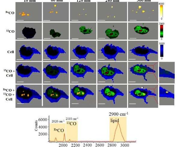
By designing special gold nanoflowers decorated with rhenium complexes (Re@Au), the team created a nanocatalyst that can reduce CO2 to CO within nerve cells. These nanostructures not only speed up the reaction but also enhance the Raman signal, allowing scientists to “see” the reaction happening in real-time.
Importantly, this was achieved using 3D Raman imaging, a non-invasive method that provides detailed, spatial insights into the chemical reactions occurring within live cells.
Their study revealed that upon light irradiation, the Re@Au nanocatalysts convert CO2 into CO with high selectivity, without harmful byproducts. Notably, the CO produced inside the cells was shown to promote neurite growth and reduce amyloid-beta protein levels — two effects relevant to treating neurodegenerative conditions like Alzheimer’s disease.
This pioneering work demonstrates how molecular reactions can be visualized in their natural cellular environment. By combining nanotechnology, photochemistry, and biomedical imaging, the study opens new doors for developing light-controlled therapies based on gas molecules like CO.
“We hope this research paves the way for future catalytic therapies that can be precisely activated inside the human body,” said Prof. Kien Voon Kong.


