


Bioengineers at Brigham and Women’s Hospital have developed a new technique to help determine if chemotherapy is working in as few as eight hours after treatment. The new approach, which can also be used for monitoring the effectiveness of immunotherapy, has shown success in pre-clinical models.
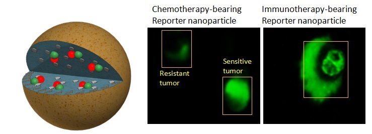
The technology utilizes a nanoparticle, carrying anti-cancer drugs, that glows green when cancer cells begin dying. Researchers, using the “reporter nanoparticles” that responds to a particular enzyme known as caspase, which is activated when cells die, were able to distinguish between a tumor that is drug-sensitive or drug-resistant much faster than conventional detection methods such as PET scans, CT and MRI. The findings were published online March 28 in the Proceedings of the National Academy of Sciences.
[pullquote]Early detection of whether or not a selected treatment is working can significantly influence the direction of treatment and improve quality of life for patients.[/pullquote]
“Using this approach, the cells light up the moment a cancer drug starts working,” co-corresponding author Shiladitya Sengupta, Ph.D., principal investigator in BWH’s Division of Bioengineering, said in a prepared statement. “We can determine if a cancer therapy is effective within hours of treatment. Our long-term goal is to find a way to monitor outcomes very early so that we don’t give a chemotherapy drug to patients who are not responding to it.”
Early detection of whether or not a selected treatment is working can significantly influence the direction of treatment and improve quality of life for patients.
In a pre-clinical model of prostate cancer, the team saw an approximately 400 percent increase in fluorescence in tumors that were sensitive to a common chemotherapeutic agent, paclitaxel, compared to tumors that were not sensitive to the drug. When the scientists separately tested an immunotherapy that targets PD-L 1 in a pre-clinical model of melanoma, after five days they also saw a significant increase in fluorescence in tumors that were treated with the therapy-loaded nanoparticles.
“We’ve demonstrated that this technique can help us directly visualize and measure the responsiveness of tumors to both types of drugs,” co-corresponding author Ashish Kulkarni, an instructor in the Division of Biomedical Engineering at BWH, said in a statement. “Current techniques, which rely on measurements of the size or metabolic state of the tumor, are sometimes unable to detect the effectiveness of an immunotherapeutic agent as the volume of the tumor may actually increase as immune cells being to flood in to attack the tumor.”
The engineered reporter nanoparticles, on the other hand, can provide an accurate measure of whether or not cancer cells are dying, Kulkarni said.
The team is actively working to design radiotracers that be used in humans, but safety and efficacy tests will need to be conducted before the technology can be converted into clinical applications.


