|
Getting your Trinity Audio player ready...
|
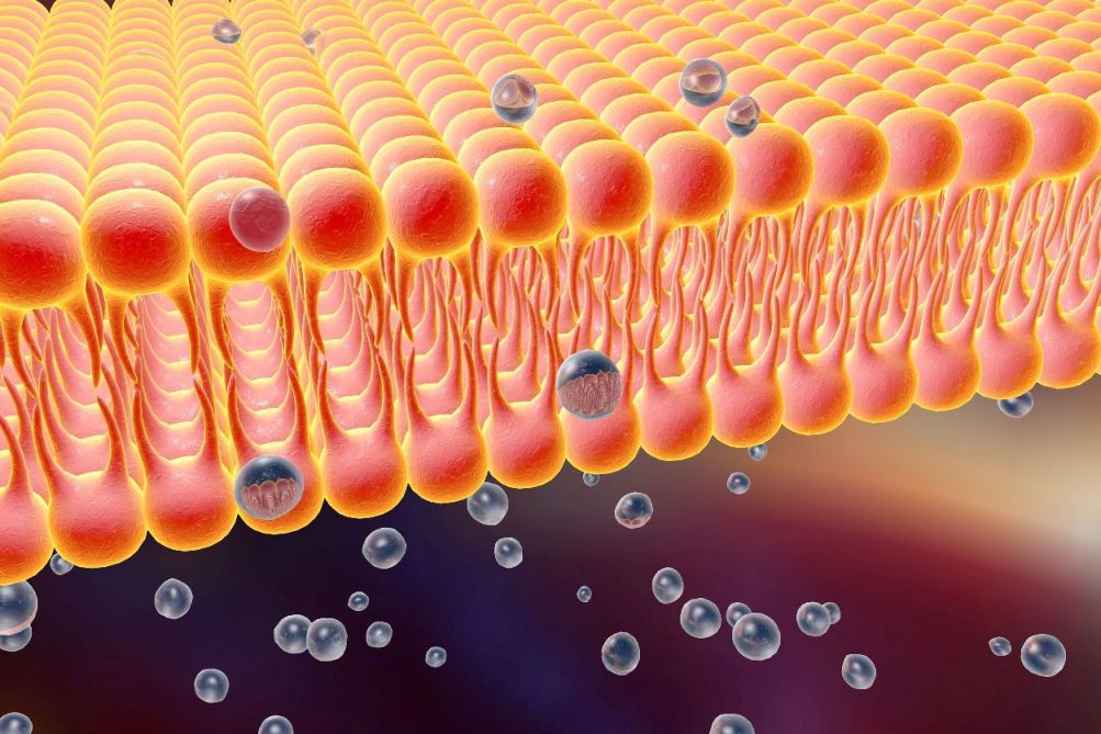
Scientists have developed new fluorescent probes that prove the existence of cell membrane structures called ‘lipid rafts’, allowing researchers to study how toxins and viruses invade cells.

Credit : Kateryna Kon / 123rf
Scientists from Japan, India and the US have observed lipid rafts in live cells for the first time. These rafts are active sections of the cell membrane responsible for signal transduction as well as the entry of toxins into cells.
The existence of lipid rafts had been assumed for over 25 years, but had never been observed in living cells.
To solve this enigma, the team focused on the behaviours of gangliosides: lipid molecules that were thought to play a central role in forming lipid rafts.
However, scientists only vaguely understood how gangliosides work because, until now, they lacked probes that could accurately track the lipids’ movements. Previous ganglioside analogues (in which florescent dye is attached) did not partition into rafts, even in artificial model systems. Researchers suspect the dyes were hydrophobic and altered how the ganglioside interacted with the cell membrane.
So, instead of just attaching a fluorescent marker to a ganglioside, the team chemically synthesized four whole gangliosides with fluorescent markers attached at specific locations. They determined which ones accurately mimicked real gangliosides, partitioning into rafts in the model system.
When the team inserted the new analogues into a living cell and used high-definition, single fluorescent-molecule imaging, they were finally able to directly document the actions of specific gangliosides in a living cell for the first time.
The researchers observed how gangliosides form lipid rafts with cholesterol and a receptor protein called CD59. It turns out these molecules interact transiently for only tens of milliseconds to form a lipid raft, and then quickly move to form a new raft. That’s why no one could observe the rafts in real cells before.
“Our findings established the concept of dynamic [lipid rafts]: their constituent molecules assemble to form [rafts], do their jobs [quickly] and then move away for the next assembly to perform the next task,” says Dr Kenichi Suzuki of Kyoto University’s Institute for Integrated Cell-Material Sciences and the paper’s co-author.
The research team next plans to use the fluorescent analogues to investigate how gangliosides regulate the activation of other receptors.


